Condition
Epiretinal membrane
Medical reviewer Mr Mohamed Mohyudin, Consultant Ophthalmic SurgeonArticle reviewed
About this review
The reviewer checked the version identified in this article’s review record.
Epiretinal membrane at a glance
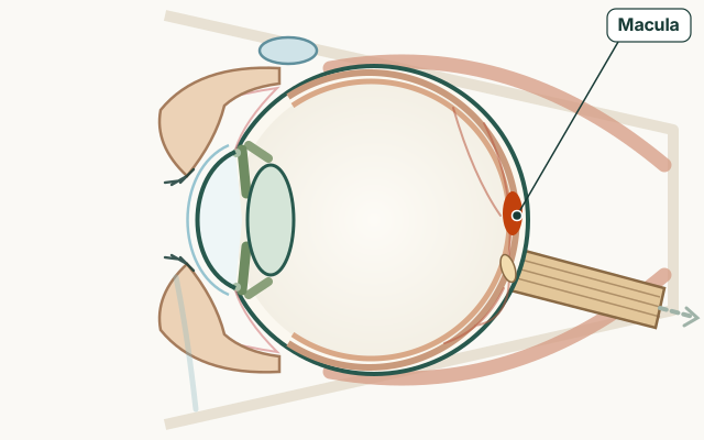
- Part of the eye
- Macula
- Common signs
- Straight lines looking wavy, bent or smaller (metamorphopsia)
- Blurred or misty central vision
- Difficulty reading small print or seeing fine detail
- A central blind spot in more advanced cases
- Treatment options
- Condition group
- Retina and optic nerve
What is an epiretinal membrane?
The retina is the light-sensing layer at the back of the eye. An epiretinal membrane (ERM) forms when cells multiply on the inner surface of the retina over the macula, gently pulling and wrinkling it. It is sometimes called macular pucker or cellophane maculopathy. It is different from a macular hole, though both affect central vision.
Symptoms of an epiretinal membrane
Symptoms depend on how much the membrane contracts.
- Straight lines looking wavy, bent or smaller (metamorphopsia)
- Blurred or misty central vision
- Difficulty reading small print or seeing fine detail
- A central blind spot in more advanced cases
- Usually painless and gradual, often in one eye
Causes and who it affects
Many ERMs have no clear cause and are found at routine eye checks in older adults. They can follow posterior vitreous detachment, retinal tears, laser treatment, inflammation, or retinal vein occlusion. Diabetes and eye injury are occasional associations. Most people with an ERM do not have another serious eye disease.
Diagnosis
Optometrists or ophthalmologists see membrane shine and retinal wrinkling during dilated examination. OCT scanning confirms thickness and how much the macula is distorted. An Amsler grid at home can help you notice new waviness between visits.
How is an epiretinal membrane treated?
Mild ERMs with little symptom impact are monitored every six to twelve months. If central vision limits reading, driving or work, vitrectomy surgery can peel the membrane. Surgery improves distortion in many cases but does not always restore normal vision; risks include cataract formation and rare complications, which your surgeon will discuss.
When to get medical advice about an epiretinal membrane
Book assessment for new central distortion or blur, especially if sudden — that can signal wet macular degeneration or other treatable problems. Stable mild distortion found at a routine test can usually be monitored on a planned schedule.
Practical guides
Treatments & Surgery
Frequently asked questions
Is an epiretinal membrane the same as a macular hole?
No. An ERM is scar tissue on the retinal surface that wrinkles the macula. A macular hole is an actual gap in central retina tissue. Both blur central vision but need different treatment approaches.
Does every epiretinal membrane need surgery?
No. Many are mild and stable. Surgery is considered when symptoms affect daily tasks and the potential benefits outweigh surgical risks.
Can an epiretinal membrane cause distorted vision?
Yes. Wavy or bent straight lines (metamorphopsia) are a hallmark symptom when the membrane pulls on the macula.
What is the surgery for epiretinal membrane?
Vitrectomy removes the vitreous gel and peels the membrane from the macular surface. It is usually a day-case procedure under local anaesthetic.
Can an epiretinal membrane get worse?
Some membranes stay stable for years; others slowly thicken and affect vision more. Regular OCT monitoring tracks change.
Sources for this information
These references support the information on this page. Follow the linked source for its full context.