Condition
Retinal artery occlusion (eye stroke)
Medical reviewer Mr Mohamed Mohyudin, Consultant Ophthalmic SurgeonArticle reviewed
About this review
The reviewer checked the version identified in this article’s review record.
Retinal artery occlusion at a glance
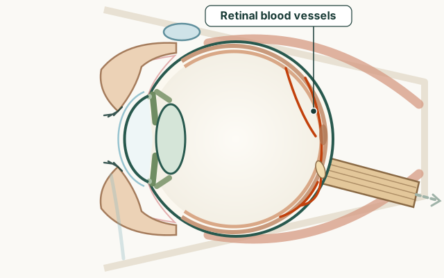
- Part of the eye
- Retinal blood vessels
- Common signs
- Sudden severe or complete vision loss in one eye
- Usually painless
- May follow brief prior episodes of temporary vision loss
- Sometimes associated with other stroke symptoms if brain is also affected
- Condition group
- Retina and optic nerve
- Medical code (ICD-10)
H34
What is retinal artery occlusion?
The retina needs continuous blood flow. When an artery is blocked by clot or embolus, retinal tissue loses oxygen within minutes and vision drops suddenly, usually without pain. Central retinal artery occlusion affects the whole retina; branch artery occlusion affects a sector of vision.
Amaurosis fugax — a warning sign
Amaurosis fugax is temporary monocular vision loss, often described as a curtain or grey-out lasting seconds to minutes, then recovery. It suggests emboli from carotid artery disease or heart rhythm problems. Treat it as a transient ischaemic event: emergency stroke assessment even if vision returns to normal.
Symptoms of retinal artery occlusion
Typical features of established artery occlusion:
- Sudden severe or complete vision loss in one eye
- Usually painless
- May follow brief prior episodes of temporary vision loss
- Sometimes associated with other stroke symptoms if brain is also affected
Causes and links
Risk factors mirror stroke: high blood pressure, diabetes, smoking, high cholesterol, atrial fibrillation, and carotid artery narrowing. Giant cell arteritis in older adults can inflame arteries supplying the eye and needs immediate steroid treatment — a different emergency with headache and jaw pain.
Treatment and urgency
Emergency eye and stroke teams assess within hours. Treatments may attempt to dislodge emboli (ocular massage, lowering eye pressure, inhaling carbon dioxide mixtures) but vision recovery is limited once retina is damaged. Essential care includes searching for stroke source, starting antiplatelet or anticoagulant therapy, and controlling vascular risk factors.
Practical guides
Frequently asked questions
What is amaurosis fugax?
It is sudden temporary vision loss in one eye, often like a curtain passing across sight. It can warn of stroke risk and needs emergency assessment even if vision returns.
Is retinal artery occlusion an emergency?
Yes. Sudden painless vision loss in one eye needs immediate hospital care for the eye and evaluation of stroke risk.
Can vision return after a retinal artery occlusion?
Significant recovery is uncommon once the retina has been starved of blood, which is why brief warning episodes and immediate care matter.
Is an eye stroke the same as a brain stroke?
It is ischaemic damage to the eye's circulation, often from the same clot sources as brain stroke. Emergency teams assess both eye and general vascular health.
What should I do if vision goes dark briefly then returns?
Seek emergency stroke assessment the same day. Do not wait for a repeat event.
Sources for this information
These references support the information on this page. Follow the linked source for its full context.