Condition
Fuchs endothelial dystrophy
Medical reviewer Mr Mohamed Mohyudin, Consultant Ophthalmic SurgeonArticle reviewed
About this review
The reviewer checked the version identified in this article’s review record.
Fuchs dystrophy at a glance
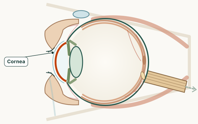
- Part of the eye
- Cornea
- Common signs
- Blurry or hazy vision, often worse on waking
- Glare and halos around lights
- Gradual difficulty with contrast and reading
- Sometimes a rough or swollen corneal surface
- Treatment options
- Condition group
- Cornea and front of eye
- Medical code (ICD-10)
H18.5
What is Fuchs dystrophy?
The inner lining of the cornea normally pumps fluid out to keep the cornea clear. In Fuchs dystrophy these cells slowly die, fluid builds up overnight, and the cornea becomes cloudy.
Symptoms of Fuchs dystrophy
Symptoms build slowly over years.
- Blurry or hazy vision, often worse on waking
- Glare and halos around lights
- Gradual difficulty with contrast and reading
- Sometimes a rough or swollen corneal surface
What causes Fuchs dystrophy?
It is usually inherited and more common in women. It is unrelated to Fuchs heterochromic uveitis. Cataract surgery can temporarily worsen swelling, so planning with a corneal specialist helps.
How is Fuchs dystrophy treated?
Early cases may use salty drops or ointment to reduce swelling. Advanced disease is treated with partial-thickness corneal transplant techniques that replace only the inner layer, with good success rates.
Treatments & Surgery
Frequently asked questions
Why is vision worse in the morning with Fuchs dystrophy?
The cornea swells overnight when the inner pump cells are weak, then may clear somewhat during the day. Morning blur is a classic pattern.
Is Fuchs dystrophy the same as cataracts?
No. Fuchs dystrophy affects the cornea's inner lining, while cataracts cloud the lens inside the eye. Both can cause glare and blur.
Can Fuchs dystrophy be cured?
Drops can help early swelling, but advanced cases are often treated successfully with modern partial corneal transplants that replace the diseased inner layer.
Sources for this information
These references support the information on this page. Follow the linked source for its full context.