Condition
Glaucoma
Medical reviewer Mr Mohamed Mohyudin, Consultant Ophthalmic SurgeonArticle reviewed
About this review
The reviewer checked the version identified in this article’s review record.
References for this section: NHS — GlaucomaNational Eye Institute - Glaucoma
Your next step →Glaucoma at a glance
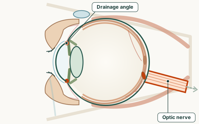
- Part of the eye
- Optic nerve and drainage angle
- Common signs
- Gradual loss of side (peripheral) vision, often unnoticed until advanced — open-angle
- Bumping into things or missing objects to one side in later stages
- Sudden severe eye pain and headache — angle-closure, an emergency
- A red eye, blurred vision and rainbow-coloured halos around lights — an emergency
- Treatment options
- Condition group
- Glaucoma and eye pressure
- Medical code (ICD-10)
H40
What is glaucoma?
Glaucoma damages the optic nerve that carries vision to the brain. Eye pressure is a major risk factor, but some people develop glaucoma with pressure in the statistically normal range (normal-tension glaucoma). Seek emergency care straight away if you have sudden severe eye pain, headache, nausea, rainbow halos and blurred vision in one eye — that pattern can be acute angle-closure glaucoma.
Open-angle glaucoma is slow and usually painless. People often notice it only when side vision is already reduced, or when an optometrist finds a suspicious nerve or field. That is why regular tests matter more than waiting for a symptom.
We do not publish a single global “success rate” for drops or surgery. Published figures vary by disease type, how late it is found, and how “success” is defined. Lost nerve fibres do not grow back.
What are the symptoms of glaucoma?
The most common type, open-angle glaucoma, usually causes no symptoms in its early stages. Vision loss starts at the edges and creeps in slowly, and the other eye often compensates, so many people only find out at a routine eye test. Acute angle-closure glaucoma is different: symptoms come on suddenly and need emergency care.
- Gradual loss of side (peripheral) vision, often unnoticed until advanced — open-angle
- Bumping into things or missing objects to one side in later stages
- Sudden severe eye pain and headache — angle-closure, an emergency
- A red eye, blurred vision and rainbow-coloured halos around lights — an emergency
- Nausea or vomiting together with eye pain — an emergency
What are the main types of glaucoma?
The names describe the drainage angle where fluid leaves the eye. In primary open-angle glaucoma the angle looks open but drainage is inefficient over years. In angle-closure the iris crowds or blocks that drain, so pressure can rise fast.
East Asian heritage raises angle-closure risk; African-Caribbean and Hispanic heritage raises open-angle risk. Family history matters for both. These are risk markers, not a diagnosis.
Two patterns — not a diagnosis from this table
| Pattern | Typical pace | Typical warning | Usual first steps after diagnosis |
|---|---|---|---|
| Primary open-angle | Years, often silent | None until side vision is damaged | Drops, selective laser, lifelong monitoring |
| Acute angle-closure | Hours | Pain, nausea, halos, red eye, blur | Emergency pressure-lowering, then laser iridotomy |
| Normal-tension | Years | Often none; nerve looks damaged despite ‘normal’ pressure | Lower pressure further; investigate other causes if needed |
Who is at risk of glaucoma, and how is it diagnosed?
Risk rises with age over 40, a first-degree relative with glaucoma, high eye pressure, short-sightedness for open-angle types, long-sightedness for angle-closure types, diabetes, long-term steroid use, and previous eye injury. Adults should have regular eye tests that can include pressure, optic-nerve assessment and visual fields when indicated.
Diagnosis is not one high pressure reading. Clinicians repeat pressure, look at the nerve (often with OCT), map the visual field, and may use gonioscopy to see the drainage angle. One isolated number does not equal glaucoma.
If you take steroid tablets, inhalers, creams around the eyes or steroid drops, say so. Steroids can raise pressure in susceptible people.
- Tonometry — measures eye pressure
- Optic nerve examination, often with OCT imaging
- Visual field test — maps side vision
- Gonioscopy — inspects the drainage angle
- Pachymetry — corneal thickness can change how pressure is interpreted
How is glaucoma treated?
Treatment lowers pressure to slow further damage. It does not reverse existing field loss. Most people start with daily drops. Selective laser trabeculoplasty (SLT) can improve drainage in open-angle disease. Laser iridotomy is used when the angle is at risk of closing. Surgery (trabeculectomy or MIGS devices) is for when drops and laser are not enough.
Missed drops allow pressure spikes. If a drop stings, blurs, or changes breathing or heart rate, tell the prescriber — alternatives exist. Do not stop a prescribed drop without talking to your eye team first.
Tell every doctor and anaesthetist you use glaucoma medicines. Some drugs interact with eye pressure or with surgery plans.
Can I drive and work with glaucoma?
Driving depends on the legal visual standard where you live — including visual fields, not only the letter chart. In the UK, glaucoma field loss can mean you must notify the licensing agency even if you still read the number plate. We do not list every country’s rule; ask your clinic and the local licensing body.
Night driving and crowded visual tasks get harder when side vision shrinks. Occupational driving and work at heights may have extra rules.
When is glaucoma an emergency?
Acute angle-closure is an emergency: severe pain, headache, nausea or vomiting, halos, a red eye and sudden blur. Same-day care also matters for sudden vision loss, a dark curtain, or a painful red eye after surgery. Having open-angle glaucoma does not rule out these emergencies.
What does sudden vision loss mean?Red eyeHalos around lights
Understand it visually
Explore the diagram
The optic nerve carries visual information from the retina to the brain. Eye examinations assess both this nerve and eye pressure.
Glaucoma damages the optic nerve. It may develop without early symptoms and can occur even when eye pressure is within the usual range.
- Lens
- Retina
- Optic nerve
Simplified educational diagram. It does not show what every person sees or replace an eye examination.
Practical guides
Treatments & Surgery
Frequently asked questions
Does glaucoma have early symptoms?
Open-angle glaucoma usually has none until side vision is damaged. Sudden pain, headache, halos and blur suggest acute angle-closure — an emergency.
Will I go blind from glaucoma?
Most people whose glaucoma is found early and who use their treatment as prescribed keep useful sight for life. Sight already lost cannot be restored, which is why regular checks and daily treatment matter.
Can glaucoma be cured?
No. Treatment can usually slow or stop further loss. Earlier detection protects more remaining sight.
How often should I use glaucoma drops?
Exactly as prescribed. Missed doses allow pressure spikes. The using glaucoma drops guide covers technique, including waiting between different bottles.
Is glaucoma hereditary?
Family history increases risk. Close adult relatives of people with glaucoma should have regular eye tests, not a one-off ‘quick look’.
What is normal-tension glaucoma?
Optic nerve damage with eye pressure in the statistically normal range. Treatment still aims to lower pressure further after other causes are considered.
Will cannabis or eye ‘exercises’ treat glaucoma?
No reliable public guidance supports either as a substitute for prescribed pressure-lowering. Do not stop drops for an unproven product.
Sources for this information
These references support the information on this page. Follow the linked source for its full context.